Are Fallen Arches Flat Feet?
Overview
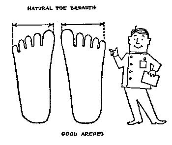
Fallen arches, also called flat feet or pes planus, is a condition in which the entire bottom of the foot touches the ground when a person is standing. A normal foot has an arch between the heel and the ball of the foot. Fallen arches are typically an acquired condition, though flat feet may be present from childhood if the arch never developed in the first place. In some cases, usually those in which the arch never developed, flat feet are not a cause for concern. If they develop in adulthood, however, causing a rigid flat foot, the condition is usually accompanied by pain and can cause serious health issues.
Causes
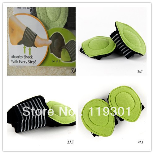
Fallen arches may be caused by a number of causes, including increased elastin during pregnancy, arthritis, injury, excessive stress on the foot, fused bones in the foot, or an extra bone. They may cause not only foot pain, but also pain in the legs, knees, and back and a loss of mobility. The condition is most often treated with orthotics, structures placed in the shoes to support the feet, but this may not be enough for severe cases. Exercises to strengthen and rebuild the arches can also be helpful. Surgery is sometimes the best method of treatment, as it can completely rebuild the arches and has lasting results, but it is quite expensive and considered a last resort.
Symptoms
Not everyone who has flat feet experiences symptoms. Others, however, feel persistent pain in their feet and wearing shoes can prove additionally painful. Others only feel symptoms when they walk more than normal, go jogging or participate in a sport that involves running or kicking.
Diagnosis
If you notice that your feet are flat, but you?re not really experiencing any pain, then you?re probably okay to go without a visit to the podiatrist (unless, of course, you have a lack of feeling in your foot). You can schedule a hair appointment instead, or maybe see a movie. However, once painful symptoms start to appear, it?s better to skip the hirsute (or cinematic) experience and go see your foot doctor. Your podiatrist will likely make the diagnosis by examining your foot visually, asking about symptoms you may be experiencing, and may test your muscle strength. You may be asked to stand on your toes (in a ballerina pose, if you prefer, although that?s certainly not required), or walk around the examining room, and you may need to show the podiatrist your shoes. He or she may comment on your excellent taste in footwear, but is more likely to check your shoes for signs of wear that may indicate fallen arches. Your podiatrist may recommend X-rays, a CT scan or an MRI in order to get a look at the interior of your foot, although the best diagnosis usually comes from the doctor?s own in-person examination.
Is flat footedness genetic?
Non Surgical Treatment
The typical treatment for pain from fallen arches is an arch insert. While many people experience dramatic pain relief from this, others continue to suffer from chronic achy feet despite the arch support. The problem with this approach is that it does not do anything to strengthen the weak ligaments that may be at the root of the problem and, thus, does not alleviate the chronic pain that people with this condition experience. Another standard practice of modern medicine is to use steroids or to prescribe anti-inflammatory medications. However, in the long run, these treatments do more damage than good. Cortisone shots and anti-inflammatory drugs have been shown to produce short-term pain benefit, but both result in long-term loss of function and even more chronic pain by actually inhibiting the healing process of soft tissues and accelerating cartilage degeneration. Plus, long-term use of these drugs can lead to other sources of chronic pain, allergies and leaky gut syndrome.
Surgical Treatment
In cases of flat feet that have progressed substantially or have failed to improve with non-surgical treatment, surgery may be required and in some advanced cases, surgery may be the only option. Your foot and ankle surgeon will determine the best approach for you.
Prevention
Sit up straight in a chair with your feet flat on the ground. Scrunch up the toes of one foot as if you are trying to grab hold of the floor then use your toes to drag your foot a small distance forwards. Do this a couple of times on each foot, but don?t use your leg muscles to push your foot forward -- the movement should come solely from the muscles in your feet. Sit in a chair and place a cleaning cloth, towel or small ball on the floor at your feet. Use the toes of one foot to grasp the object and lift it off the floor. This action will require you to clench your toes and contract your arch. Once you have lifted the object a little way off the floor, try to throw it in the air and catch it by stretching your toes and arch out and upwards. Repeat the exercise several times on both feet. Sit on the floor with your legs straight out in front of you then bend your knees out to either side and place the soles of your feet together so your legs form a diamond. Hold on to your ankles and, keeping your heels together at all times, separate your feet so your toes point out to either side. Open and close your feet in this way several times, making sure your little toes stay in contact with the floor throughout the exercise. Starting in the same position, try separating your heels, keeping your toes together at all times.
After Care
Time off work depends on the type of work as well as the surgical procedures performed. . A patient will be required to be non-weight bearing in a cast or splint and use crutches for four to twelve weeks. Usually a patient can return to work in one to two weeks if they are able to work while seated. If a person's job requires standing and walking, return to work may take several weeks. Complete recovery may take six months to a full year. Complications can occur as with all surgeries, but are minimized by strictly following your surgeon's post-operative instructions. The main complications include infection, bone that is slow to heal or does not heal, progression or reoccurrence of deformity, a stiff foot, and the need for further surgery. Many of the above complications can be avoided by only putting weight on the operative foot when allowed by your surgeon.
Fallen arches, also called flat feet or pes planus, is a condition in which the entire bottom of the foot touches the ground when a person is standing. A normal foot has an arch between the heel and the ball of the foot. Fallen arches are typically an acquired condition, though flat feet may be present from childhood if the arch never developed in the first place. In some cases, usually those in which the arch never developed, flat feet are not a cause for concern. If they develop in adulthood, however, causing a rigid flat foot, the condition is usually accompanied by pain and can cause serious health issues.
Causes
Fallen arches may be caused by a number of causes, including increased elastin during pregnancy, arthritis, injury, excessive stress on the foot, fused bones in the foot, or an extra bone. They may cause not only foot pain, but also pain in the legs, knees, and back and a loss of mobility. The condition is most often treated with orthotics, structures placed in the shoes to support the feet, but this may not be enough for severe cases. Exercises to strengthen and rebuild the arches can also be helpful. Surgery is sometimes the best method of treatment, as it can completely rebuild the arches and has lasting results, but it is quite expensive and considered a last resort.
Symptoms
Not everyone who has flat feet experiences symptoms. Others, however, feel persistent pain in their feet and wearing shoes can prove additionally painful. Others only feel symptoms when they walk more than normal, go jogging or participate in a sport that involves running or kicking.
Diagnosis
If you notice that your feet are flat, but you?re not really experiencing any pain, then you?re probably okay to go without a visit to the podiatrist (unless, of course, you have a lack of feeling in your foot). You can schedule a hair appointment instead, or maybe see a movie. However, once painful symptoms start to appear, it?s better to skip the hirsute (or cinematic) experience and go see your foot doctor. Your podiatrist will likely make the diagnosis by examining your foot visually, asking about symptoms you may be experiencing, and may test your muscle strength. You may be asked to stand on your toes (in a ballerina pose, if you prefer, although that?s certainly not required), or walk around the examining room, and you may need to show the podiatrist your shoes. He or she may comment on your excellent taste in footwear, but is more likely to check your shoes for signs of wear that may indicate fallen arches. Your podiatrist may recommend X-rays, a CT scan or an MRI in order to get a look at the interior of your foot, although the best diagnosis usually comes from the doctor?s own in-person examination.
Is flat footedness genetic?
Non Surgical Treatment
The typical treatment for pain from fallen arches is an arch insert. While many people experience dramatic pain relief from this, others continue to suffer from chronic achy feet despite the arch support. The problem with this approach is that it does not do anything to strengthen the weak ligaments that may be at the root of the problem and, thus, does not alleviate the chronic pain that people with this condition experience. Another standard practice of modern medicine is to use steroids or to prescribe anti-inflammatory medications. However, in the long run, these treatments do more damage than good. Cortisone shots and anti-inflammatory drugs have been shown to produce short-term pain benefit, but both result in long-term loss of function and even more chronic pain by actually inhibiting the healing process of soft tissues and accelerating cartilage degeneration. Plus, long-term use of these drugs can lead to other sources of chronic pain, allergies and leaky gut syndrome.
Surgical Treatment
In cases of flat feet that have progressed substantially or have failed to improve with non-surgical treatment, surgery may be required and in some advanced cases, surgery may be the only option. Your foot and ankle surgeon will determine the best approach for you.
Prevention
Sit up straight in a chair with your feet flat on the ground. Scrunch up the toes of one foot as if you are trying to grab hold of the floor then use your toes to drag your foot a small distance forwards. Do this a couple of times on each foot, but don?t use your leg muscles to push your foot forward -- the movement should come solely from the muscles in your feet. Sit in a chair and place a cleaning cloth, towel or small ball on the floor at your feet. Use the toes of one foot to grasp the object and lift it off the floor. This action will require you to clench your toes and contract your arch. Once you have lifted the object a little way off the floor, try to throw it in the air and catch it by stretching your toes and arch out and upwards. Repeat the exercise several times on both feet. Sit on the floor with your legs straight out in front of you then bend your knees out to either side and place the soles of your feet together so your legs form a diamond. Hold on to your ankles and, keeping your heels together at all times, separate your feet so your toes point out to either side. Open and close your feet in this way several times, making sure your little toes stay in contact with the floor throughout the exercise. Starting in the same position, try separating your heels, keeping your toes together at all times.
After Care
Time off work depends on the type of work as well as the surgical procedures performed. . A patient will be required to be non-weight bearing in a cast or splint and use crutches for four to twelve weeks. Usually a patient can return to work in one to two weeks if they are able to work while seated. If a person's job requires standing and walking, return to work may take several weeks. Complete recovery may take six months to a full year. Complications can occur as with all surgeries, but are minimized by strictly following your surgeon's post-operative instructions. The main complications include infection, bone that is slow to heal or does not heal, progression or reoccurrence of deformity, a stiff foot, and the need for further surgery. Many of the above complications can be avoided by only putting weight on the operative foot when allowed by your surgeon.
Heel Aches
Overview
Heel pain is a very challenging problem as it can be local and/or referred. It has been more prevalent recently due to the hard grounds on which people have to run. Commonly people will present with heel pain, thrusting an x-ray at you, and being adamant that the problem is a ?heel spur?. This is defined as a small bone that grows from the heel, directing forwards towards the toes. This may be as small as 1 mm to anything as large as 8 - 10 mm. Most of the time, this is an incidental finding, as there many heels that are pain free that have heel spurs evident on x-rays. The spur is thought to be a result of traction of the plantar fascia on the heel. In some cases, the spur may contribute to the symptoms, but is not the main cause. This should be explained very carefully to the patient, as the focus on the spur may limit the recovery, as the patient may believe that the only way to eliminate the pain is to remove the spur.
Causes
There are many reasons why people experience heel pain. Based on what we see in our office, heel pain affects, probably more than one in every four people. A lot of this is caused from conditions within the foot. These conditions could be related to hyper pronation, which is where you get a collapse of the foot or even a high arch of the foot called cavus foot. The underlying cause is something internal, within the bone structure. These problems are usually something you will have all your life. Hyper pronation is a hereditary issue where you can get an under development of a particular bone, usually in the ankle, and it causes a dislocation or a misalignment of the ankle on the heel. It throws off, not only the foot with the bones, joints and ligaments of the foot, but also the bones in the ankle. It affects the internal rotation of the knee, hip, back and causes issues within those areas as well. Hyper pronation is a pretty common, but very under diagnosed condition.
Symptoms
The heel can be painful in many different ways, depending on the cause. Plantar fasciitis commonly causes intense heel pain along the bottom of the foot during the first few steps after getting out of bed in the morning. This heel pain often goes away once you start to walk around, but it may return in the late afternoon or evening. Although X-ray evidence suggests that about 10% of the general population has heels spurs, many of these people do not have any symptoms. In others, heel spurs cause pain and tenderness on the undersurface of the heel that worsen over several months. In a child, this condition causes pain and tenderness at the lower back portion of the heel. The affected heel is often sore to the touch but not obviously swollen. Bursitis involving the heel causes pain in the middle of the undersurface of the heel that worsens with prolonged standing and pain at the back of the heel that worsens if you bend your foot up or down. Pump bump, this condition causes a painful enlargement at the back of the heel, especially when wearing shoes that press against the back of the heel. Heel bruises, like bruises elsewhere in the body, may cause pain, mild swelling, soreness and a black-and-blue discoloration of the skin. Achilles tendonitis, this condition causes pain at the back of the heel where the Achilles tendon attaches to the heel. The pain typically becomes worse if you exercise or play sports, and it often is followed by soreness, stiffness and mild swelling. A trapped nerve can cause pain, numbness or tingling almost anywhere at the back, inside or undersurface of the heel. In addition, there are often other symptoms, such as swelling or discoloration - if the trapped nerve was caused by a sprain, fracture or other injury.
Diagnosis
Your GP or podiatrist (a healthcare professional who specialises in foot care) may be able to diagnose the cause of your heel pain by asking about your symptoms and examining your heel and foot. You will usually only need further tests if you have additional symptoms that suggest the cause of your heel pain is not inflammation, such as numbness or a tingling sensation in your foot - this could be a sign of nerve damage in your feet and legs (peripheral neuropathy), your foot feels hot and you have a high temperature (fever) of 38C (100.4F) or above - these could be signs of a bone infection, you have stiffness and swelling in your heel - this could be a sign of arthritis. Possible further tests may include, blood tests, X-rays - where small doses of radiation are used to detect problems with your bones and tissues, a magnetic resonance imaging (MRI) scan or ultrasound scan, which are more detailed scans.
Non Surgical Treatment
As heel pain is basically a stress problem in the tissues of the heel, the main treatment is to reduce stress. Your doctor will advise you about weight loss and appropriate footwear. A soft heel pad is useful to wear in your shoe to act as a shock-absorber when you walk. If you have a stiff ankle or tight Achilles tendon a physiotherapist can advise on exercises for these. Stretching the Achilles tendon and plantar fascia is very effective general treatment for many patients. If you have a high-arched or flat foot, a podiatrist may advise an insole to reduce stress. Simple pain-killers such as paracetamol or anti-inflammatory medicines can help reduce the pain. Ask advice from your doctor or pharmacist before taking anti-inflammatory medicines as they can have troublesome side-effects in some people. The simple measures above will help the majority of people with heel pain. If the pain continues, a splint to wear on your ankle at night to prevent your Achilles tendon tightening up while you are asleep is often very effective in improving the severe pain that many people get first thing in the morning and breaking the cycle of pain. Your GP or an orthopaedic foot and ankle surgeon or rheumatologist may inject some steroid into the attachment of the plantar fascia to damp down the inflammation. These measures will reduce the pain in most people who are not helped by simple treatment. If you still have pain after one or two injections, your doctor may want to investigate your problem a bit further. If no other medical problem or cause of stress in your heel is found, a number of other treatments can be tried. Further physiotherapy, wearing a plaster cast to rest the inflamed tissues, pain control treatments such as transcutaneous nerve stimulation (TENS) or acupuncture. Only if all non-surgical treatments fail would an operation be considered.
Surgical Treatment
It is rare to need an operation for heel pain. It would only be offered if all simpler treatments have failed and, in particular, you are a reasonable weight for your height and the stresses on your heel cannot be improved by modifying your activities or footwear. The aim of an operation is to release part of the plantar fascia from the heel bone and reduce the tension in it. Many surgeons would also explore and free the small nerves on the inner side of your heel as these are sometimes trapped by bands of tight tissue. This sort of surgery can be done through a cut about 3cm long on the inner side of your heel. Recently there has been a lot of interest in doing the operation by keyhole surgery, but this has not yet been proven to be effective and safe. Most people who have an operation are better afterwards, but it can take months to get the benefit of the operation and the wound can take a while to heal fully. Tingling or numbness on the side of the heel may occur after operation.
how to add a strap to high heels
Prevention

Being overweight can place excess pressure and strain on your feet, particularly on your heels. Losing weight, and maintaining a healthy weight by combining regular exercise with a healthy, balanced diet, can be beneficial for your feet. Wearing appropriate footwear is also important. Ideally, you should wear shoes with a low to moderate heel that supports and cushions your arches and heels. Avoid wearing shoes with no heels.
Heel pain is a very challenging problem as it can be local and/or referred. It has been more prevalent recently due to the hard grounds on which people have to run. Commonly people will present with heel pain, thrusting an x-ray at you, and being adamant that the problem is a ?heel spur?. This is defined as a small bone that grows from the heel, directing forwards towards the toes. This may be as small as 1 mm to anything as large as 8 - 10 mm. Most of the time, this is an incidental finding, as there many heels that are pain free that have heel spurs evident on x-rays. The spur is thought to be a result of traction of the plantar fascia on the heel. In some cases, the spur may contribute to the symptoms, but is not the main cause. This should be explained very carefully to the patient, as the focus on the spur may limit the recovery, as the patient may believe that the only way to eliminate the pain is to remove the spur.
Causes
There are many reasons why people experience heel pain. Based on what we see in our office, heel pain affects, probably more than one in every four people. A lot of this is caused from conditions within the foot. These conditions could be related to hyper pronation, which is where you get a collapse of the foot or even a high arch of the foot called cavus foot. The underlying cause is something internal, within the bone structure. These problems are usually something you will have all your life. Hyper pronation is a hereditary issue where you can get an under development of a particular bone, usually in the ankle, and it causes a dislocation or a misalignment of the ankle on the heel. It throws off, not only the foot with the bones, joints and ligaments of the foot, but also the bones in the ankle. It affects the internal rotation of the knee, hip, back and causes issues within those areas as well. Hyper pronation is a pretty common, but very under diagnosed condition.
Symptoms
The heel can be painful in many different ways, depending on the cause. Plantar fasciitis commonly causes intense heel pain along the bottom of the foot during the first few steps after getting out of bed in the morning. This heel pain often goes away once you start to walk around, but it may return in the late afternoon or evening. Although X-ray evidence suggests that about 10% of the general population has heels spurs, many of these people do not have any symptoms. In others, heel spurs cause pain and tenderness on the undersurface of the heel that worsen over several months. In a child, this condition causes pain and tenderness at the lower back portion of the heel. The affected heel is often sore to the touch but not obviously swollen. Bursitis involving the heel causes pain in the middle of the undersurface of the heel that worsens with prolonged standing and pain at the back of the heel that worsens if you bend your foot up or down. Pump bump, this condition causes a painful enlargement at the back of the heel, especially when wearing shoes that press against the back of the heel. Heel bruises, like bruises elsewhere in the body, may cause pain, mild swelling, soreness and a black-and-blue discoloration of the skin. Achilles tendonitis, this condition causes pain at the back of the heel where the Achilles tendon attaches to the heel. The pain typically becomes worse if you exercise or play sports, and it often is followed by soreness, stiffness and mild swelling. A trapped nerve can cause pain, numbness or tingling almost anywhere at the back, inside or undersurface of the heel. In addition, there are often other symptoms, such as swelling or discoloration - if the trapped nerve was caused by a sprain, fracture or other injury.
Diagnosis
Your GP or podiatrist (a healthcare professional who specialises in foot care) may be able to diagnose the cause of your heel pain by asking about your symptoms and examining your heel and foot. You will usually only need further tests if you have additional symptoms that suggest the cause of your heel pain is not inflammation, such as numbness or a tingling sensation in your foot - this could be a sign of nerve damage in your feet and legs (peripheral neuropathy), your foot feels hot and you have a high temperature (fever) of 38C (100.4F) or above - these could be signs of a bone infection, you have stiffness and swelling in your heel - this could be a sign of arthritis. Possible further tests may include, blood tests, X-rays - where small doses of radiation are used to detect problems with your bones and tissues, a magnetic resonance imaging (MRI) scan or ultrasound scan, which are more detailed scans.
Non Surgical Treatment
As heel pain is basically a stress problem in the tissues of the heel, the main treatment is to reduce stress. Your doctor will advise you about weight loss and appropriate footwear. A soft heel pad is useful to wear in your shoe to act as a shock-absorber when you walk. If you have a stiff ankle or tight Achilles tendon a physiotherapist can advise on exercises for these. Stretching the Achilles tendon and plantar fascia is very effective general treatment for many patients. If you have a high-arched or flat foot, a podiatrist may advise an insole to reduce stress. Simple pain-killers such as paracetamol or anti-inflammatory medicines can help reduce the pain. Ask advice from your doctor or pharmacist before taking anti-inflammatory medicines as they can have troublesome side-effects in some people. The simple measures above will help the majority of people with heel pain. If the pain continues, a splint to wear on your ankle at night to prevent your Achilles tendon tightening up while you are asleep is often very effective in improving the severe pain that many people get first thing in the morning and breaking the cycle of pain. Your GP or an orthopaedic foot and ankle surgeon or rheumatologist may inject some steroid into the attachment of the plantar fascia to damp down the inflammation. These measures will reduce the pain in most people who are not helped by simple treatment. If you still have pain after one or two injections, your doctor may want to investigate your problem a bit further. If no other medical problem or cause of stress in your heel is found, a number of other treatments can be tried. Further physiotherapy, wearing a plaster cast to rest the inflamed tissues, pain control treatments such as transcutaneous nerve stimulation (TENS) or acupuncture. Only if all non-surgical treatments fail would an operation be considered.
Surgical Treatment
It is rare to need an operation for heel pain. It would only be offered if all simpler treatments have failed and, in particular, you are a reasonable weight for your height and the stresses on your heel cannot be improved by modifying your activities or footwear. The aim of an operation is to release part of the plantar fascia from the heel bone and reduce the tension in it. Many surgeons would also explore and free the small nerves on the inner side of your heel as these are sometimes trapped by bands of tight tissue. This sort of surgery can be done through a cut about 3cm long on the inner side of your heel. Recently there has been a lot of interest in doing the operation by keyhole surgery, but this has not yet been proven to be effective and safe. Most people who have an operation are better afterwards, but it can take months to get the benefit of the operation and the wound can take a while to heal fully. Tingling or numbness on the side of the heel may occur after operation.
how to add a strap to high heels
Prevention
Being overweight can place excess pressure and strain on your feet, particularly on your heels. Losing weight, and maintaining a healthy weight by combining regular exercise with a healthy, balanced diet, can be beneficial for your feet. Wearing appropriate footwear is also important. Ideally, you should wear shoes with a low to moderate heel that supports and cushions your arches and heels. Avoid wearing shoes with no heels.
Leg Length Discrepancy Women
Overview
The two major categorizations of LLD are structural and functional. A third more minor category is environmental. In structural LLD there is an actual anatomical difference in the bones of the lower extremities where one side becomes shorter than the other. This type of LLD may be genetic, where the person is born in this way. In other cases it may be due to injury or infection through the growth phases of early childhood or adolescence. Some spinal abnormalities like scoliosis can also cause this condition. Functional LLD is where the bones are not the cause of difference but a muscle or pelvic condition has the effect of weakening the leg on one side. Conditions that can cause this are muscle inflexibility, adduction contractures and pelvic obliquity (amongst others). The third less severe category of environmental LLD is caused by discrepancies in the surface that the feet and legs are resting or walking on. Banked, uneven or curved surfaces can all cause environmental LLD. In LLD the asymmetric nature of the legs in relation to hips and back caused the centre of gravity to shift from its natural position. This then results in the body attempting to compensate by either tilting the pelvic areas towards the shorter side, increased knee flexing on the longer side, flexion of the ankle plantar and foot supination towards the shorter side.
Causes
Some children are born with absence or underdeveloped bones in the lower limbs e.g., congenital hemimelia. Others have a condition called hemihypertrophy that causes one side of the body to grow faster than the other. Sometimes, increased blood flow to one limb (as in a hemangioma or blood vessel tumor) stimulates growth to the limb. In other cases, injury or infection involving the epiphyseal plate (growth plate) of the femur or tibia inhibits or stops altogether the growth of the bone. Fractures healing in an overlapped position, even if the epiphyseal plate is not involved, can also cause limb length discrepancy. Neuromuscular problems like polio can also cause profound discrepancies, but thankfully, uncommon. Lastly, Wilms? tumor of the kidney in a child can cause hypertrophy of the lower limb on the same side. It is therefore important in a young child with hemihypertrophy to have an abdominal ultrasound exam done to rule out Wilms? tumor. It is important to distinguish true leg length discrepancy from apparent leg length discrepancy. Apparent discrepancy is due to an instability of the hip, that allows the proximal femur to migrate proximally, or due to an adduction or abduction contracture of the hip that causes pelvic obliquity, so that one hip is higher than the other. When the patient stands, it gives the impression of leg length discrepancy, when the problem is actually in the hip.
Symptoms
The symptoms of limb deformity can range from a mild difference in the appearance of a leg or arm to major loss of function of the use of an extremity. For instance, you may notice that your child has a significant limp. If there is deformity in the extremity, the patient may develop arthritis as he or she gets older, especially if the lower extremities are involved. Patients often present due to the appearance of the extremity (it looks different from the other side).
Diagnosis
The most accurate method to identify leg (limb) length inequality (discrepancy) is through radiography. It?s also the best way to differentiate an anatomical from a functional limb length inequality. Radiography, A single exposure of the standing subject, imaging the entire lower extremity. Limitations are an inherent inaccuracy in patients with hip or knee flexion contracture and the technique is subject to a magnification error. Computed Tomography (CT-scan), It has no greater accuracy compared to the standard radiography. The increased cost for CT-scan may not be justified, unless a contracture of the knee or hip has been identified or radiation exposure must be minimized. However, radiography has to be performed by a specialist, takes more time and is costly. It should only be used when accuracy is critical. Therefore two general clinical methods were developed for assessing LLI. Direct methods involve measuring limb length with a tape measure between 2 defined points, in stand. Two common points are the anterior iliac spine and the medial malleolus or the anterior inferior iliac spine and lateral malleolus. Be careful, however, because there is a great deal of criticism and debate surrounds the accuracy of tape measure methods. If you choose for this method, keep following topics and possible errors in mind. Always use the mean of at least 2 or 3 measures. If possible, compare measures between 2 or more clinicians. Iliac asymmetries may mask or accentuate a limb length inequality. Unilateral deviations in the long axis of the lower limb (eg. Genu varum,?) may mask or accentuate a limb length inequality. Asymmetrical position of the umbilicus. Joint contractures. Indirect methods. Palpation of bony landmarks, most commonly the iliac crests or anterior iliac spines, in stand. These methods consist in detecting if bony landmarks are at (horizontal) level or if limb length inequality is present. Palpation and visual estimation of the iliac crest (or SIAS) in combination with the use of blocks or book pages of known thickness under the shorter limb to adjust the level of the iliac crests (or SIAS) appears to be the best (most accurate and precise) clinical method to asses limb inequality. You should keep in mind that asymmetric pelvic rotations in planes other than the frontal plane may be associated with limb length inequality. A review of the literature suggest, therefore, that the greater trochanter major and as many pelvic landmarks should be palpated and compared (left trochanter with right trochanter) when the block correction method is used.
Non Surgical Treatment
Structural leg length discrepancy can be treated with a heel lift in the shorter leg?s shoe, if the leg length is greater than 5 mm. The use and size of the heel lift is determined by a physical therapist based on how much lift is needed to restore proper lumbopelvic biomechanics. In certain cases, surgical intervention may be needed to either shorten or lengthen the limb. An important component to any surgical procedure to correct leg length discrepancies is physical therapy. Physical therapy helps to stretch muscles and maintain joint flexibility, which is essential in the healing process. For a functional leg length discrepancy no heel lift is required, but proper manual therapy techniques and specific therapeutic exercise is needed to treat and normalize pelvic and lower extremity compensations. The number of treatments needed to hold the pelvis in a symmetrical position is different for each patient based on their presentation and biomechanical dysfunctions in their lower back, pelvis, hip, knee, and foot/ankle. If you have pain in your lower back or lower extremity and possibly a length discrepancy; the two symptoms could be related. A good place to start would be a physical therapy evaluation to determine whether you have a leg length discrepancy and if it could be contributing to your lower back pain, hip pain, knee pain, or leg pain.

what can i do to get taller?
Surgical Treatment
The type of surgery depends on the type of problem. Outpatient procedures may be used to alter the growth of the limb. This is often done through small incisions. If an outpatient procedure is done, your child can continue with most regular activities. Other times, surgery may be very involved and require the use of an external device that is attached to the limb with pins and wires. This device may be left on for months to correct the deformity or lengthen the leg. If this type of surgery is required, your child will be making weekly visits to Cincinnati Children's.
The two major categorizations of LLD are structural and functional. A third more minor category is environmental. In structural LLD there is an actual anatomical difference in the bones of the lower extremities where one side becomes shorter than the other. This type of LLD may be genetic, where the person is born in this way. In other cases it may be due to injury or infection through the growth phases of early childhood or adolescence. Some spinal abnormalities like scoliosis can also cause this condition. Functional LLD is where the bones are not the cause of difference but a muscle or pelvic condition has the effect of weakening the leg on one side. Conditions that can cause this are muscle inflexibility, adduction contractures and pelvic obliquity (amongst others). The third less severe category of environmental LLD is caused by discrepancies in the surface that the feet and legs are resting or walking on. Banked, uneven or curved surfaces can all cause environmental LLD. In LLD the asymmetric nature of the legs in relation to hips and back caused the centre of gravity to shift from its natural position. This then results in the body attempting to compensate by either tilting the pelvic areas towards the shorter side, increased knee flexing on the longer side, flexion of the ankle plantar and foot supination towards the shorter side.
Causes
Some children are born with absence or underdeveloped bones in the lower limbs e.g., congenital hemimelia. Others have a condition called hemihypertrophy that causes one side of the body to grow faster than the other. Sometimes, increased blood flow to one limb (as in a hemangioma or blood vessel tumor) stimulates growth to the limb. In other cases, injury or infection involving the epiphyseal plate (growth plate) of the femur or tibia inhibits or stops altogether the growth of the bone. Fractures healing in an overlapped position, even if the epiphyseal plate is not involved, can also cause limb length discrepancy. Neuromuscular problems like polio can also cause profound discrepancies, but thankfully, uncommon. Lastly, Wilms? tumor of the kidney in a child can cause hypertrophy of the lower limb on the same side. It is therefore important in a young child with hemihypertrophy to have an abdominal ultrasound exam done to rule out Wilms? tumor. It is important to distinguish true leg length discrepancy from apparent leg length discrepancy. Apparent discrepancy is due to an instability of the hip, that allows the proximal femur to migrate proximally, or due to an adduction or abduction contracture of the hip that causes pelvic obliquity, so that one hip is higher than the other. When the patient stands, it gives the impression of leg length discrepancy, when the problem is actually in the hip.
Symptoms
The symptoms of limb deformity can range from a mild difference in the appearance of a leg or arm to major loss of function of the use of an extremity. For instance, you may notice that your child has a significant limp. If there is deformity in the extremity, the patient may develop arthritis as he or she gets older, especially if the lower extremities are involved. Patients often present due to the appearance of the extremity (it looks different from the other side).
Diagnosis
The most accurate method to identify leg (limb) length inequality (discrepancy) is through radiography. It?s also the best way to differentiate an anatomical from a functional limb length inequality. Radiography, A single exposure of the standing subject, imaging the entire lower extremity. Limitations are an inherent inaccuracy in patients with hip or knee flexion contracture and the technique is subject to a magnification error. Computed Tomography (CT-scan), It has no greater accuracy compared to the standard radiography. The increased cost for CT-scan may not be justified, unless a contracture of the knee or hip has been identified or radiation exposure must be minimized. However, radiography has to be performed by a specialist, takes more time and is costly. It should only be used when accuracy is critical. Therefore two general clinical methods were developed for assessing LLI. Direct methods involve measuring limb length with a tape measure between 2 defined points, in stand. Two common points are the anterior iliac spine and the medial malleolus or the anterior inferior iliac spine and lateral malleolus. Be careful, however, because there is a great deal of criticism and debate surrounds the accuracy of tape measure methods. If you choose for this method, keep following topics and possible errors in mind. Always use the mean of at least 2 or 3 measures. If possible, compare measures between 2 or more clinicians. Iliac asymmetries may mask or accentuate a limb length inequality. Unilateral deviations in the long axis of the lower limb (eg. Genu varum,?) may mask or accentuate a limb length inequality. Asymmetrical position of the umbilicus. Joint contractures. Indirect methods. Palpation of bony landmarks, most commonly the iliac crests or anterior iliac spines, in stand. These methods consist in detecting if bony landmarks are at (horizontal) level or if limb length inequality is present. Palpation and visual estimation of the iliac crest (or SIAS) in combination with the use of blocks or book pages of known thickness under the shorter limb to adjust the level of the iliac crests (or SIAS) appears to be the best (most accurate and precise) clinical method to asses limb inequality. You should keep in mind that asymmetric pelvic rotations in planes other than the frontal plane may be associated with limb length inequality. A review of the literature suggest, therefore, that the greater trochanter major and as many pelvic landmarks should be palpated and compared (left trochanter with right trochanter) when the block correction method is used.
Non Surgical Treatment
Structural leg length discrepancy can be treated with a heel lift in the shorter leg?s shoe, if the leg length is greater than 5 mm. The use and size of the heel lift is determined by a physical therapist based on how much lift is needed to restore proper lumbopelvic biomechanics. In certain cases, surgical intervention may be needed to either shorten or lengthen the limb. An important component to any surgical procedure to correct leg length discrepancies is physical therapy. Physical therapy helps to stretch muscles and maintain joint flexibility, which is essential in the healing process. For a functional leg length discrepancy no heel lift is required, but proper manual therapy techniques and specific therapeutic exercise is needed to treat and normalize pelvic and lower extremity compensations. The number of treatments needed to hold the pelvis in a symmetrical position is different for each patient based on their presentation and biomechanical dysfunctions in their lower back, pelvis, hip, knee, and foot/ankle. If you have pain in your lower back or lower extremity and possibly a length discrepancy; the two symptoms could be related. A good place to start would be a physical therapy evaluation to determine whether you have a leg length discrepancy and if it could be contributing to your lower back pain, hip pain, knee pain, or leg pain.
what can i do to get taller?
Surgical Treatment
The type of surgery depends on the type of problem. Outpatient procedures may be used to alter the growth of the limb. This is often done through small incisions. If an outpatient procedure is done, your child can continue with most regular activities. Other times, surgery may be very involved and require the use of an external device that is attached to the limb with pins and wires. This device may be left on for months to correct the deformity or lengthen the leg. If this type of surgery is required, your child will be making weekly visits to Cincinnati Children's.
What Causes Mortons Neuroma
Overview
 Morton's neuroma is an inflammation of the nerves in the foot that go to the toes. Although the name includes the word ?neuroma,? it is not really a tumor. It can affect any of the toes in the foot. However, it most often affects the nerves that run between the third and fourth, or second and third toes.
Causes
The cause of this problem is often due to impingement of the plantar nerve fibres between the metatarsal heads and the intermetatarsal ligament. It is entirely a biomechanical phenomenon. Differential diagnoses include stress fracture, capsulitis, bursitis or ligament injury at the metatarsal-phalangeal joint, a tendon sheath ganglion, foreign-body reaction and nerve-sheath tumour.
Symptoms
Morton's neuroma can cause a very painful burning or sharp pain in your foot that feels worse when you walk. It may feel like a small lump inside the ball of your foot. It is usually between the third and fourth toes, but it can also be between other toes.
Diagnosis
A doctor can usually identify Morton's neuroma during a physical exam. He or she will squeeze or press on the bottom of your foot or squeeze your toes together to see if it hurts. Your doctor may also order an X-ray of your foot to make sure nothing else is causing the pain.
Non Surgical Treatment
The first line of treatment is to try modifying footwear. Often simply wearing broader fitting shoes can reduce pressure on the neuroma and so reduce pain. Orthotic inserts can also help as they can again help reduce pressure on certain parts of the foot. Padding and taping the toe area is another option. In some cases a steroid injection into the foot may be suggested. This can be done as a day case without the need for anaesthesia and helps reduce inflation of the nerve. It can halt the pain in round 70 % of cases. Sometimes a combination of alcohol and local anaesthesia may be injected as this helps reduce pain.
Surgical Treatment
Surgery. This is the last and most permanent course of action. This surgery is used as a last resort as it often comes with a series of side affects including the risk of making the pain worse. This surgery can be performed by Orthopedic surgeons as well as Podiatric surgeons.
Morton's neuroma is an inflammation of the nerves in the foot that go to the toes. Although the name includes the word ?neuroma,? it is not really a tumor. It can affect any of the toes in the foot. However, it most often affects the nerves that run between the third and fourth, or second and third toes.Causes
The cause of this problem is often due to impingement of the plantar nerve fibres between the metatarsal heads and the intermetatarsal ligament. It is entirely a biomechanical phenomenon. Differential diagnoses include stress fracture, capsulitis, bursitis or ligament injury at the metatarsal-phalangeal joint, a tendon sheath ganglion, foreign-body reaction and nerve-sheath tumour.
Symptoms
Morton's neuroma can cause a very painful burning or sharp pain in your foot that feels worse when you walk. It may feel like a small lump inside the ball of your foot. It is usually between the third and fourth toes, but it can also be between other toes.
Diagnosis
A doctor can usually identify Morton's neuroma during a physical exam. He or she will squeeze or press on the bottom of your foot or squeeze your toes together to see if it hurts. Your doctor may also order an X-ray of your foot to make sure nothing else is causing the pain.
Non Surgical Treatment
The first line of treatment is to try modifying footwear. Often simply wearing broader fitting shoes can reduce pressure on the neuroma and so reduce pain. Orthotic inserts can also help as they can again help reduce pressure on certain parts of the foot. Padding and taping the toe area is another option. In some cases a steroid injection into the foot may be suggested. This can be done as a day case without the need for anaesthesia and helps reduce inflation of the nerve. It can halt the pain in round 70 % of cases. Sometimes a combination of alcohol and local anaesthesia may be injected as this helps reduce pain.
Surgical Treatment
Surgery. This is the last and most permanent course of action. This surgery is used as a last resort as it often comes with a series of side affects including the risk of making the pain worse. This surgery can be performed by Orthopedic surgeons as well as Podiatric surgeons.
How Shoe Lifts Cure Leg Length Imbalances
There are two different types of leg length discrepancies, congenital and acquired. Congenital implies that you are born with it. One leg is anatomically shorter than the other. As a result of developmental phases of aging, the human brain senses the step pattern and recognizes some variation. The entire body usually adapts by tilting one shoulder to the "short" side. A difference of less than a quarter inch isn't blatantly excessive, doesn't need Shoe Lifts to compensate and in most cases doesn't have a serious effect over a lifetime.

Leg length inequality goes mainly undiscovered on a daily basis, yet this issue is easily fixed, and can eradicate a number of instances of chronic back pain.
Treatment for leg length inequality typically involves Shoe Lifts. These are typically low cost, commonly costing below twenty dollars, compared to a custom orthotic of $200 or maybe more. Differences over a quarter inch can take their toll on the spine and should probably be compensated for with a heel lift. In some cases, the shortage can be so extreme that it requires a full lift to both the heel and sole of the shoe.
Mid back pain is easily the most common health problem affecting men and women today. Over 80 million men and women have problems with back pain at some point in their life. It's a problem that costs employers millions annually on account of lost time and production. Fresh and better treatment solutions are constantly sought after in the hope of reducing the economical influence this issue causes.

Men and women from all corners of the earth suffer the pain of foot ache due to leg length discrepancy. In these types of situations Shoe Lifts can be of beneficial. The lifts are capable of relieving any pain in the feet. Shoe Lifts are recommended by many professional orthopaedic physicians.
So that you can support the human body in a healthy and balanced fashion, feet have got a vital task to play. Irrespective of that, it's often the most neglected region in the human body. Many people have flat-feet which means there is unequal force exerted on the feet. This causes other body parts like knees, ankles and backs to be impacted too. Shoe Lifts make sure that appropriate posture and balance are restored.
Leg length inequality goes mainly undiscovered on a daily basis, yet this issue is easily fixed, and can eradicate a number of instances of chronic back pain.
Treatment for leg length inequality typically involves Shoe Lifts. These are typically low cost, commonly costing below twenty dollars, compared to a custom orthotic of $200 or maybe more. Differences over a quarter inch can take their toll on the spine and should probably be compensated for with a heel lift. In some cases, the shortage can be so extreme that it requires a full lift to both the heel and sole of the shoe.
Mid back pain is easily the most common health problem affecting men and women today. Over 80 million men and women have problems with back pain at some point in their life. It's a problem that costs employers millions annually on account of lost time and production. Fresh and better treatment solutions are constantly sought after in the hope of reducing the economical influence this issue causes.
Men and women from all corners of the earth suffer the pain of foot ache due to leg length discrepancy. In these types of situations Shoe Lifts can be of beneficial. The lifts are capable of relieving any pain in the feet. Shoe Lifts are recommended by many professional orthopaedic physicians.
So that you can support the human body in a healthy and balanced fashion, feet have got a vital task to play. Irrespective of that, it's often the most neglected region in the human body. Many people have flat-feet which means there is unequal force exerted on the feet. This causes other body parts like knees, ankles and backs to be impacted too. Shoe Lifts make sure that appropriate posture and balance are restored.
The Answer To Leg Length Imbalances Are Shoe Lifts
There are not one but two different kinds of leg length discrepancies, congenital and acquired. Congenital indicates that you are born with it. One leg is structurally shorter in comparison to the other. Through developmental periods of aging, the human brain picks up on the step pattern and recognizes some difference. The body usually adapts by dipping one shoulder to the "short" side. A difference of less than a quarter inch is not grossly uncommon, does not need Shoe Lifts to compensate and generally does not have a serious effect over a lifetime.

Leg length inequality goes mainly undiscovered on a daily basis, yet this problem is easily corrected, and can eliminate quite a few cases of back pain.
Therapy for leg length inequality typically involves Shoe Lifts. These are typically low cost, ordinarily being less than twenty dollars, compared to a custom orthotic of $200 and up. When the amount of leg length inequality begins to exceed half an inch, a whole sole lift is generally the better choice than a heel lift. This prevents the foot from being unnecessarily stressed in an abnormal position.
Upper back pain is the most prevalent health problem impacting men and women today. Around 80 million people have problems with back pain at some stage in their life. It is a problem that costs companies millions of dollars every year due to time lost and output. New and more effective treatment solutions are always sought after in the hope of lowering economic impact this condition causes.

People from all corners of the earth suffer from foot ache due to leg length discrepancy. In these types of cases Shoe Lifts are usually of very helpful. The lifts are capable of alleviating any discomfort in the feet. Shoe Lifts are recommended by numerous professional orthopaedic orthopedists.
In order to support the body in a healthy and balanced fashion, feet have a crucial job to play. Irrespective of that, it is often the most overlooked area in the human body. Many people have flat-feet meaning there may be unequal force placed on the feet. This causes other parts of the body like knees, ankles and backs to be impacted too. Shoe Lifts ensure that the right posture and balance are restored.
Leg length inequality goes mainly undiscovered on a daily basis, yet this problem is easily corrected, and can eliminate quite a few cases of back pain.
Therapy for leg length inequality typically involves Shoe Lifts. These are typically low cost, ordinarily being less than twenty dollars, compared to a custom orthotic of $200 and up. When the amount of leg length inequality begins to exceed half an inch, a whole sole lift is generally the better choice than a heel lift. This prevents the foot from being unnecessarily stressed in an abnormal position.
Upper back pain is the most prevalent health problem impacting men and women today. Around 80 million people have problems with back pain at some stage in their life. It is a problem that costs companies millions of dollars every year due to time lost and output. New and more effective treatment solutions are always sought after in the hope of lowering economic impact this condition causes.
People from all corners of the earth suffer from foot ache due to leg length discrepancy. In these types of cases Shoe Lifts are usually of very helpful. The lifts are capable of alleviating any discomfort in the feet. Shoe Lifts are recommended by numerous professional orthopaedic orthopedists.
In order to support the body in a healthy and balanced fashion, feet have a crucial job to play. Irrespective of that, it is often the most overlooked area in the human body. Many people have flat-feet meaning there may be unequal force placed on the feet. This causes other parts of the body like knees, ankles and backs to be impacted too. Shoe Lifts ensure that the right posture and balance are restored.
Shoe Lifts The Chiropodists Choice For Leg Length Discrepancy
There are actually not one but two different kinds of leg length discrepancies, congenital and acquired. Congenital means you are born with it. One leg is anatomically shorter in comparison to the other. Through developmental phases of aging, the brain picks up on the stride pattern and identifies some variance. Our bodies usually adapts by tilting one shoulder over to the "short" side. A difference of less than a quarter inch is not grossly irregular, demand Shoe Lifts to compensate and normally won't have a serious effect over a lifetime.
Leg length inequality goes typically undiagnosed on a daily basis, yet this issue is simply fixed, and can eradicate numerous incidents of back ache.
Therapy for leg length inequality usually consists of Shoe Lifts. These are cost-effective, regularly costing below twenty dollars, compared to a custom orthotic of $200 plus. When the amount of leg length inequality begins to exceed half an inch, a whole sole lift is generally the better choice than a heel lift. This prevents the foot from being unnecessarily stressed in an abnormal position.
Back pain is easily the most common health problem affecting men and women today. Over 80 million people are afflicted by back pain at some stage in their life. It's a problem which costs companies millions of dollars yearly because of lost time and output. Fresh and superior treatment solutions are continually sought after in the hope of lowering economical impact this condition causes.
Men and women from all corners of the world experience foot ache as a result of leg length discrepancy. In most of these cases Shoe Lifts might be of very beneficial. The lifts are capable of alleviating any discomfort and pain in the feet. Shoe Lifts are recommended by countless specialist orthopaedic practitioners".
To be able to support the body in a well-balanced manner, your feet have a vital part to play. Despite that, it is often the most overlooked region of the human body. Many people have flat-feet which means there may be unequal force exerted on the feet. This causes other body parts like knees, ankles and backs to be impacted too. Shoe Lifts guarantee that suitable posture and balance are restored.